

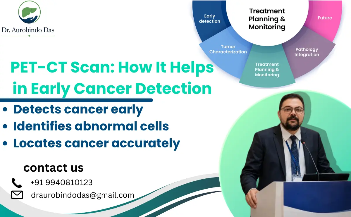
Cancer diagnosis can be stressful, especially when symptoms are unclear or when doctors need to understand whether an abnormal area is cancerous, localized, or spreading. Modern imaging has made this evaluation more precise. Among the advanced investigations available today, PET-CT combines functional information from positron emission tomography with detailed anatomical images from computed tomography.
A PET-CT Scan for Early Cancer Detection can help doctors identify areas with increased metabolic activity, locate suspicious lesions, assess the extent of disease, and evaluate how a cancer responds to treatment. Unlike imaging that primarily shows anatomy, PET imaging can provide information about how tissues are behaving at a cellular or metabolic level.
This guide explains how PET-CT Scan for Early Cancer Detection works, who may need it, how to prepare, what happens during the procedure, its benefits and limitations, and what patients can expect afterward.
Understanding PET-CT and Its Role in Cancer Care
What is a PET-CT scan?
A PET-CT scan combines two imaging techniques during one examination. CT creates detailed anatomical images showing the size, shape, and location of structures. PET provides functional information by showing areas where a radiotracer accumulates. Combining both sets of information allows doctors to understand both where an abnormality is located and how metabolically active it may be.
The most commonly used PET tracer in cancer imaging is fluorodeoxyglucose, or FDG. FDG behaves similarly to glucose. Many cancer cells use glucose at a higher rate than surrounding normal tissues, so areas of increased uptake may become visible on the PET portion of the examination.
This is one reason PET-CT Scan for Early Cancer Detection can provide information that may not be available from an anatomical scan alone. By combining metabolic and structural information, doctors can obtain a more comprehensive picture of suspicious areas.
Why is PET-CT used in cancer?
PET-CT may be recommended for several reasons, including:
- Investigating a suspected cancer
- Determining the extent or stage of an established cancer
- Looking for cancer that has spread to other parts of the body
- Evaluating whether treatment is working
- Checking for possible recurrence after treatment
- Helping doctors plan appropriate treatment
The exact role varies according to the cancer type. PET-CT is particularly valuable when metabolic information can influence staging or treatment decisions.
In selected patients, PET-CT Scan for Early Cancer Detection may provide additional information that helps doctors decide whether further investigations, biopsy, surgery, or another treatment approach is appropriate.
Symptoms that may lead to further cancer evaluation
PET-CT itself does not diagnose a specific symptom. A doctor may consider advanced imaging when symptoms or previous investigations raise concern about an underlying disease.
Possible warning symptoms can include:
- Unexplained weight loss
- Persistent abdominal pain
- A new or growing lump
- Persistent cough or breathing difficulty
- Unexplained bleeding
- Changes in bowel or bladder habits
- Difficulty swallowing
- Persistent fatigue
- Recurrent fever without an obvious cause
- Jaundice or unexplained changes in liver function
These symptoms can occur for many non-cancerous reasons. Having one or more of them does not mean that a person has cancer. However, persistent or unexplained symptoms should be medically evaluated rather than ignored.
Causes and risk factors
Cancer develops through complex changes in cells, and there is usually no single cause. Risk factors vary according to the type of cancer and can include tobacco exposure, alcohol consumption, certain infections, inherited genetic changes, obesity, environmental exposures, age, and family history.
A PET-CT scan does not identify the original cause of cancer. Instead, it can provide important information about abnormal tissue activity and the distribution of disease when clinically indicated.
PET-CT Scan for Early Cancer Detection: Diagnosis and Procedure
How is cancer evaluated before PET-CT?
The diagnostic pathway usually begins with a medical consultation. Depending on the symptoms, a doctor may recommend blood tests, ultrasound, endoscopy, CT, MRI, mammography, or other investigations.
If an abnormality is detected or cancer is strongly suspected, PET-CT may be recommended to answer a specific clinical question. Importantly, PET-CT findings are interpreted together with the patient’s symptoms, examination, laboratory results, previous imaging, and—when required—pathology.
A PET-CT scan can show increased metabolic activity, but increased uptake is not automatically cancer. Inflammation and infection can also produce increased FDG uptake. Therefore, suspicious findings may require additional testing or biopsy.
For appropriate patients, PET-CT Scan for Early Cancer Detection can form an important part of a broader diagnostic pathway rather than being used as an isolated test.
How does the PET-CT procedure work?
The procedure generally follows these steps:
1. Preparation
Patients are commonly instructed not to eat for several hours before an FDG PET-CT, although water is usually permitted. Specific instructions can vary according to the protocol and the patient’s medical condition. Patients should inform the medical team about medications, diabetes, allergies, pregnancy or possible pregnancy, and breastfeeding.
2. Radiotracer injection
A small amount of radioactive radiotracer is administered through an intravenous injection. The patient then rests quietly while the tracer circulates and accumulates in tissues.
3. Uptake period
The tracer commonly requires approximately 30–60 minutes to distribute through the body. Patients are generally asked to remain relaxed and avoid unnecessary movement or talking during this period.
4. Imaging
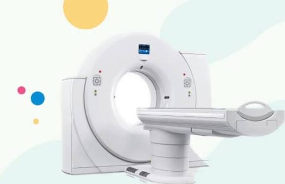
The patient lies on a scanning table that moves through the PET-CT machine. The CT component provides anatomical information, while PET records metabolic activity.
5. Image interpretation
A trained radiologist or nuclear medicine physician interprets the images and prepares a report for the treating doctor.
How does PET-CT support early cancer detection?
The major strength of PET imaging is that it can reveal metabolic changes that may occur before structural abnormalities become obvious on some other imaging examinations. However, this does not mean that PET-CT can reliably detect every cancer at its earliest stage.
The usefulness of PET-CT Scan for Early Cancer Detection depends on the cancer type, tumor biology, tumor size, and tracer used. Some cancers may show low FDG uptake, while certain benign inflammatory conditions can appear metabolically active.
Therefore, PET-CT should be viewed as an important part of selected cancer evaluation rather than a universal cancer screening test.
Is PET-CT a replacement for biopsy?
No. Imaging can identify suspicious areas and help determine disease distribution, but a biopsy is often required to establish a definitive tissue diagnosis.
PET-CT can also help doctors identify appropriate areas for further evaluation and understand whether disease may have spread. Treatment decisions are made using the complete clinical picture rather than the PET-CT result alone.
For this reason, PET-CT Scan for Early Cancer Detection should always be interpreted alongside clinical assessment and other diagnostic findings.
Benefits, Comparison, Recovery and Treatment Planning
Benefits of PET-CT in cancer management
A PET-CT Scan for Early Cancer Detection can offer several important advantages when appropriately indicated:
- Combines functional and anatomical information
- Helps identify metabolically active abnormalities
- Can help determine the extent of cancer
- May identify distant disease that changes treatment planning
- Can help assess response to therapy
- Can help investigate suspected recurrence
- Provides whole-body information in many cancer protocols
- May reduce the need for multiple separate imaging examinations in selected situations
Because PET and CT are performed together, doctors can correlate metabolic activity with precise anatomical locations.
PET-CT vs CT or MRI
CT, MRI, ultrasound, and PET-CT each have different strengths.
CT: Provides detailed anatomical images and is widely used for evaluating tumors, organs, lymph nodes, and other structures.
MRI: Provides excellent soft-tissue contrast and is especially useful for selected neurological, pelvic, musculoskeletal, liver, and other conditions.
PET-CT: Adds metabolic information to anatomical imaging and can be particularly helpful for staging, treatment assessment, and detecting recurrent or metastatic disease in appropriate cancers.
The choice is not necessarily about which test is “better.” The appropriate examination depends on the clinical question. In some cases, several imaging techniques are complementary.
When doctors are considering PET-CT Scan for Early Cancer Detection, they assess the patient’s medical history, symptoms, suspected cancer type, previous test results, and the specific information required from imaging.
What happens after PET-CT?
Most patients can return to normal activities relatively quickly after the examination unless their treating team provides different instructions. The radioactive tracer naturally loses radioactivity over time and is also eliminated from the body, including through urine.
Drinking adequate water after the scan may help with elimination, unless fluid intake is restricted for another medical reason. Patients should follow the imaging center’s specific post-scan instructions.
Risks and limitations
PET-CT uses ionizing radiation from both the radiotracer and CT component. The radiation exposure is considered acceptable when the examination is appropriately justified, but unnecessary imaging should be avoided.
Other limitations include false-positive findings from inflammation or infection and false-negative findings in some tumors with low metabolic activity. Blood glucose levels can also influence FDG PET imaging, making appropriate preparation particularly important for patients with diabetes.
PET-CT is therefore most useful when ordered for a clear clinical reason and interpreted by appropriately trained specialists.
Treatment after PET-CT
PET-CT does not itself treat cancer. Instead, the information it provides can influence the next stage of care.
Depending on the diagnosis and stage, treatment may involve surgery, chemotherapy, radiation therapy, targeted therapy, immunotherapy, ablation, interventional procedures, or combinations of these approaches.
For gastrointestinal, liver, pancreatic, and biliary cancers, PET-CT findings may contribute to multidisciplinary treatment planning. Surgical decisions can depend on tumor location, vascular involvement, lymph-node status, distant spread, liver function, and the patient’s overall health.
In these situations, PET-CT Scan for Early Cancer Detection may provide useful information that supports a more individualized approach to cancer management.
Frequently Asked Questions and Expert Care
1. Can a PET-CT scan detect cancer at an early stage?
A PET-CT Scan for Early Cancer Detection can identify metabolic abnormalities that may sometimes appear before structural changes are obvious on other imaging. However, its ability to detect early cancer depends on the cancer type, tumor size, biological behavior, and tracer used. It is not a universal screening test for every cancer.
2. Does PET-CT confirm cancer?
Not always. Increased tracer uptake can occur with cancer, but inflammation and infection can also cause uptake. A biopsy may be necessary to confirm cancer.
3. Is PET-CT painful?
The scan itself is generally painless. The main discomfort is usually the intravenous injection used to administer the radiotracer.
4. How long does a PET-CT take?
The exact duration varies. The tracer commonly requires around 30–60 minutes for uptake, followed by the imaging portion.
5. Can I eat before PET-CT?
Patients undergoing FDG PET-CT are commonly asked to fast for several hours, but preparation instructions vary. Always follow the instructions provided by the imaging center.
6. Can PET-CT show whether cancer has spread?
Yes. One important use of PET-CT is evaluating whether cancer has spread to lymph nodes or distant organs.
7. Is PET-CT useful after cancer treatment?
Yes. In selected cancers, it may help doctors evaluate treatment response or investigate possible recurrence.
8. Is PET-CT safe?
PET-CT involves radiation exposure, but the examination is generally considered safe when medically indicated. Patients should inform their doctor if they are pregnant, may be pregnant, or are breastfeeding.
9. Should everyone get a PET-CT for cancer screening?
No. PET-CT is not recommended as a routine whole-body screening examination for everyone. Doctors recommend imaging based on individual risk, symptoms, examination findings, previous tests, and the suspected disease.
About Dr. Sri Aurobindo Prasad Das
Dr. Sri Aurobindo Prasad Das, also known as Dr. Aurobindo Das, is an Associate Consultant in the Institute of Surgical Gastroenterology, GI and HPB Onco-Surgery and Liver Transplantation at Sir Ganga Ram Hospital, New Delhi. His qualifications include MBBS from JIPMER, MS from JIPMER, MCh in GI Surgery from AIIMS, DNB in GI Surgery, MNAMS, FAIS, and FMAS.
His areas of interest include liver transplantation, laparoscopic GI surgery, and complex hepatobiliary-pancreatic (HPB) surgery. He has also been associated with JIPMER, AIIMS, Fortis Noida, and Apollo Hospitals Chennai for training and professional experience.
As a gastrointestinal and HPB surgeon, Dr. Aurobindo Das provides specialist care for complex diseases involving the digestive system, liver, pancreas, gallbladder, and bile ducts. His clinical services include surgical gastroenterology, complex HPB surgery, laparoscopic GI procedures, and liver transplantation.
The Institute of Surgical Gastroenterology, GI and HPB Onco-Surgery and Liver Transplantation at Sir Ganga Ram Hospital provides specialist services for complex gastrointestinal, hepatobiliary, pancreatic, oncological, and transplant conditions.
For patients undergoing cancer evaluation, specialist consultation can help determine whether imaging such as PET-CT is appropriate and how the findings should be integrated into the overall treatment plan.
Conclusion
A PET-CT Scan for Early Cancer Detection can be a valuable tool in modern oncology when used for the right patient and the right clinical question. By combining metabolic information from PET with detailed anatomical information from CT, the examination can help doctors evaluate suspicious disease, determine cancer spread, monitor treatment response, and investigate recurrence.
At the same time, patients should understand that PET-CT is not a substitute for clinical evaluation or biopsy when tissue confirmation is required. An abnormal PET result does not automatically mean cancer, and a normal result does not exclude every possible malignancy.
If you have persistent symptoms, an abnormal imaging report, a known cancer diagnosis, or concerns about possible disease spread, discuss your situation with an experienced specialist. Appropriate imaging and timely evaluation can help doctors make more informed decisions about the next step in care.
For patients seeking specialist gastrointestinal, HPB, liver, and cancer-related surgical evaluation in New Delhi, consultation with Dr. Sri Aurobindo Prasad Das (Dr. Aurobindo Das) at Sir Ganga Ram Hospital can help establish an individualized diagnostic and treatment pathway.
Book an appointment for expert consultation and discuss whether a PET-CT examination is appropriate for your condition.
Dr. Sri Aurobindo Prasad Das (Dr. Aurobindo Das)
Gastrointestinal & HPB Surgical Care | Liver Transplantation
Sir Ganga Ram Hospital
Near Karol Bagh Metro, Old Rajendra Nagar, New Delhi – 110060
Phone: 9940810123
Email: draurobindodas@gmail.com